HOME > Weekly Case > Weekly Case
Weekly Case
종결
Case No. 141
0000-00-00~0000-00-00
출제자 : 관리자
Hit : 296
| Period | 0000-00-00~0000-00-00 |
|---|---|
| Diagnosis | |
| Clinical information | 39세 여자, tinnitus로 내원. |
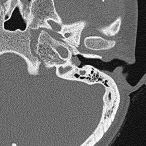
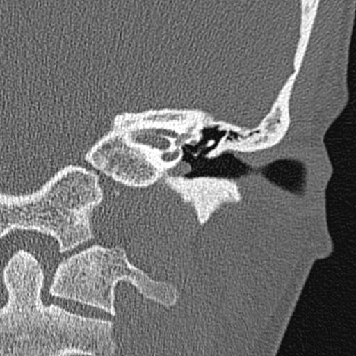
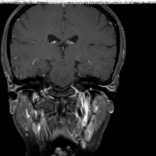
| Discussion | Glomus Tympanicum 영상소견 Temporal bone CT axial, coronal image에서 soft tissue denstiy를 보이는 mass가 promontory에 인접한 hypotympanum에 보이며 bone erosion은 보이지 않음. MRI상 middle ear cavity에 small mass lesion이 보이며 강한 조영증강을 보임. Review 정의: benign tumor arising from glomus bodies situated on cochlear promontory 위치: Cochlear promontory (a rounded hollow prominence, fromed by the projection outward of the first turn of the cochlear). CN9 의 Extracranial branch인 tympanic br.(jacobsen nerve)가 middle ear sensation 을 담당하며 Inf. Tympanic canalicus 를 통해 middle ear 로 진입. Cochlear promontory 에서 tympanic plexus를 형성 영상소견 CT: Focal mass with flat base on cochlear promontory Large size인 경우 medial wall of middle ear 및 ossicle의 erosion 를 보일수 있다. MR: T1 강조영상에서 cochlear promontory에 tissue intenstiy를 보이며 T2 강조영상에서 obstructive fluid 보다 낮은 신호강도를 보이며 조영증강시 조영증강을 보인다. Differential diagnosis 1. Abberant internal carotid artery 2. Dehiscent jugular bulb 3. Congenital cholesteatoma of middle ear 4. Glomus jugulotympanicum paraganglioma 5. Facial nerve schwannoma, tympanic segment |
| Correct answer |
Correct AnswerSemi-Correct Answer |