HOME > Weekly Case > Weekly Case
Weekly Case
종결
Case No. 140
0000-00-00~0000-00-00
출제자 : 관리자
Hit : 313
| Period | 0000-00-00~0000-00-00 |
|---|---|
| Diagnosis | |
| Clinical information | 49세 남자, 2개월 간의 neck pain으로 내원. |
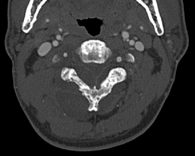
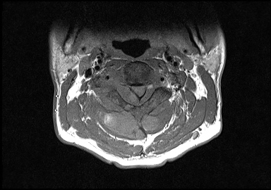
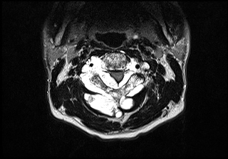
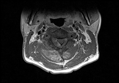
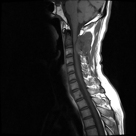
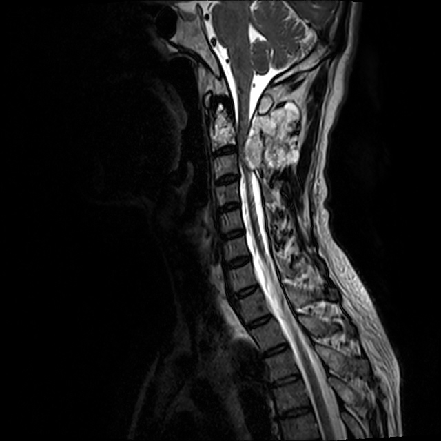
| Discussion | 진단명: Chordoma Imaging findings MR상 C2 posterior arch를 침범하고 주위 soft tissue mass를 형성하며 spinal canal내와C2 body도 같이 침범하는 mass로서 T2WI상 high signal, T1WI상 iso 및 mild enhancement를 보임. CT상 osteolytic 및 sclerotic change와 함께 mild expansion을 보임. Review - arise from fetal notochord remnant - typically involve clivus and sacrococcygeal region, rare in C-spine - vertebral body involve, perivertebral and epidural extension - sometimes main mass in epidural space-extraosseous notochordal remnant > - erosion and lytic bone destruction with remodeling on CT - typically hyperintense on T2 in most cases - heterogeneous mild enhancement - 본 증례는 vertebra의 posterior arch를 주로 involve한 점에서 unusual하나, T2WI상 very high signal을 보이고 enhance가 mild한 점 등에서 chordoma의 가능성을 생각할 수 있는 증례로 생각됨. Reference) AJR 1999;172:1423-1426 |
| Correct answer |
Correct AnswerSemi-Correct Answer |