HOME > Weekly Case > Weekly Case
Weekly Case
종결
Case No. 138
0000-00-00~0000-00-00
출제자 : 관리자
Hit : 362
| Period | 0000-00-00~0000-00-00 |
|---|---|
| Diagnosis | |
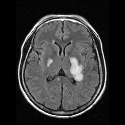
| Clinical information | 58세 여자, 3주 전부터 시작된 right side sensory change로 내원 |
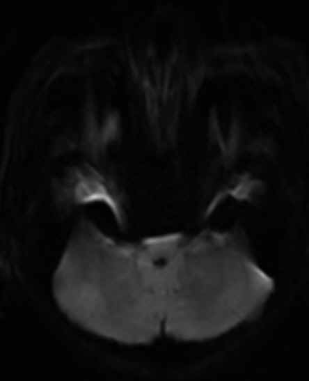
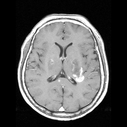
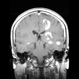
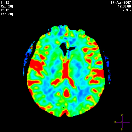
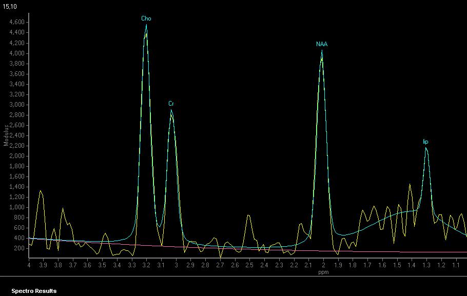
| Discussion | 진단명: Multiple sclerosis 처음 발병시 tumor 또는 infarction의심되었으나 그 후 MR상 병변이 rapid change보이고optic neuritis 발견되었으며 CSF상 IgG index증가 등의 소견 보여 multiple sclerosis로 진단됨. 추적검사상 병변 작아짐. Review of Tumefactive Multiple sclerosis MS의 일반적인 MR 영상소견은 1. Multiple white matter lesions, predominantly in the periventricular white matter 2. combined supratentorial and infratentorial lesions 3. minimal edema and mass effect 으로 정리할 수 있다. Tumefactive MS의 경우 tumor와의 감별이 어려우나 enhance 소견이나 advanced MR imaging 이 도움이 될 수 있다. 본 환자의 경우 - enhanced MR상 부분적으로 incomplete ring enhancement를 보였는데 이는 demyelinating의 특징적 소견 중 하나이다. - DWI상 high signal은 demyelinating disease의 acute stage에 보일 수 있는 소견임. - enhance되는 병변임에도 perfusion의 증가가 없거나 작은 경우 high grade tumor보다는 non-tumorous condition을 favor함. - MRS상 NAA가 약간 감소되고 Cho/Cr ratio가 약간 증가되며 Lipid/lactate peak의 증가 등의 소견도 high grade tumor보다는 non-tumorous condition을 favor함. - 그러나, 보고에 따라서는 Cho/Cr ratio가 high grade tumor와 유사하게 나오는 경우도 있어 conventional MR소견을 바탕으로 DWI, PWI, MRS 소견을 부가적으로 이용하여 진단에 도움을 받을 수 있는 것으로 생각됨. |
| Correct answer |
Correct AnswerSemi-Correct Answer |