HOME > Weekly Case > Weekly Case
Weekly Case
종결
Case No. 133
0000-00-00~0000-00-00
출제자 : 관리자
Hit : 349
| Period | 0000-00-00~0000-00-00 |
|---|---|
| Diagnosis | |
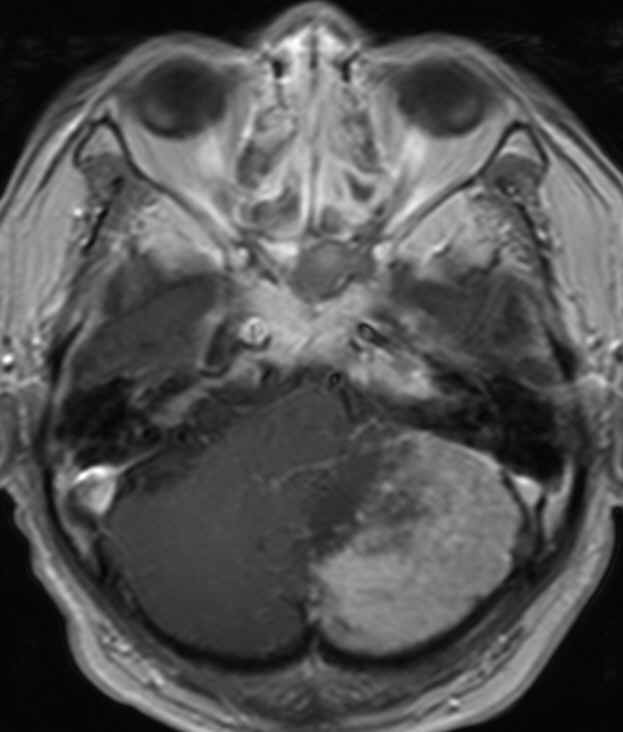
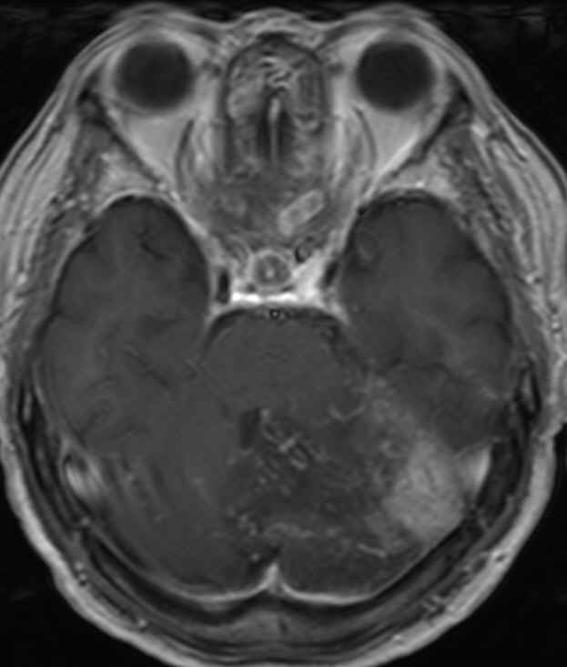
| Clinical information | 76세 남자, dizziness와 dysarthria로 내원 |
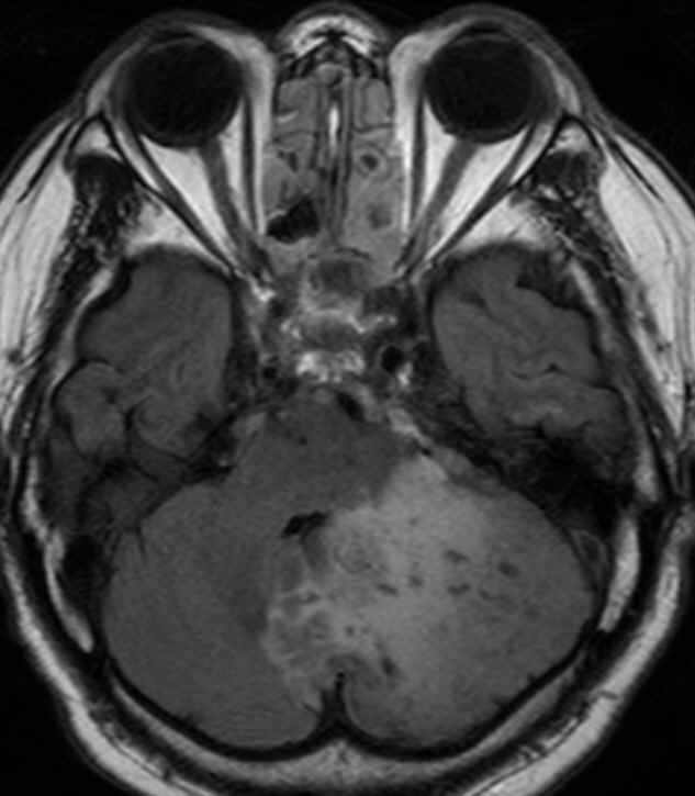
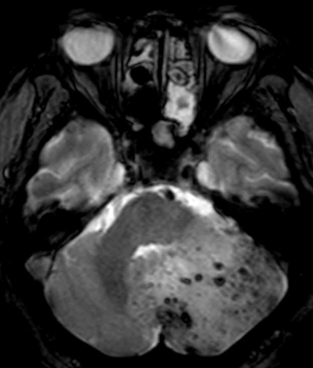
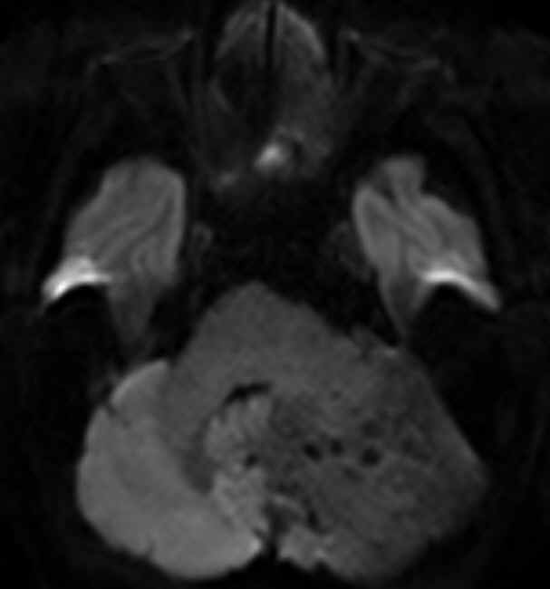
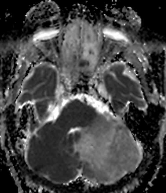
| Discussion | BRAIN MRI -Left cerebellar hemisphere에 mass effect를 갖는 FLAIR에서 high signal intensity lesion이 보이며 gradient echo image에서는 내부에 multiple hemorrhagic foci가 관찰됨. ADC map에서 이 lesion은 high signal intensity를 보이고 비교적 homogenous enhancement를 보이며 주위로 venous structure가 multiple하게 관찰됨. Left vertebral artery angiogram에서 posterior meningeal artery의 hypertrophy와 posterior meningeal artery와 직접 연결된 AV fistula가 관찰되며 이 AV fistula는 superior vermian vein과 inferior vermian vein으로 drain 되고 있음. 진단: Venous infarction due to dural AVF Dural arteriovenous fistula (DAVF) an abnormal arteriovenous shunt that occurs in the dura matter within or near a dural sinus. Tissue hypoperfusion induced by venous hypertension can cause brain ischemia and even infarction. Clinical symptoms also correlated well with the areas of impaired hemodynamic and metabolic parameters. DAVF with retrograde leptomeningeal venous drainage into a single venous sinus is more prone to brain tissue ischemia or infarction due to long-term impaired venous drainage. DWI is very useful for detecting cytotoxic edema in venous infarction but is not as territorial in distribution as arterial infarction. Reference : J Neuroimaging 2006;16:69-72. |
| Correct answer |
Correct AnswerSemi-Correct Answer |