HOME > Weekly Case > Weekly Case
Weekly Case
종결
Case No. 131
0000-00-00~0000-00-00
출제자 : 관리자
Hit : 306
| Period | 0000-00-00~0000-00-00 |
|---|---|
| Diagnosis | |
| Clinical information | 35세 남자, 3개월 전부터 시작된 diplopia로 내원. |
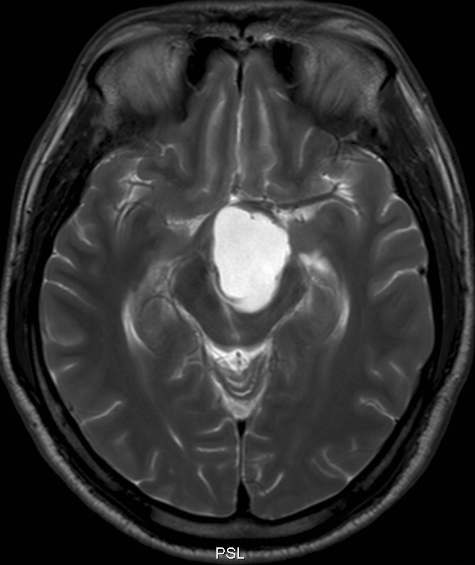
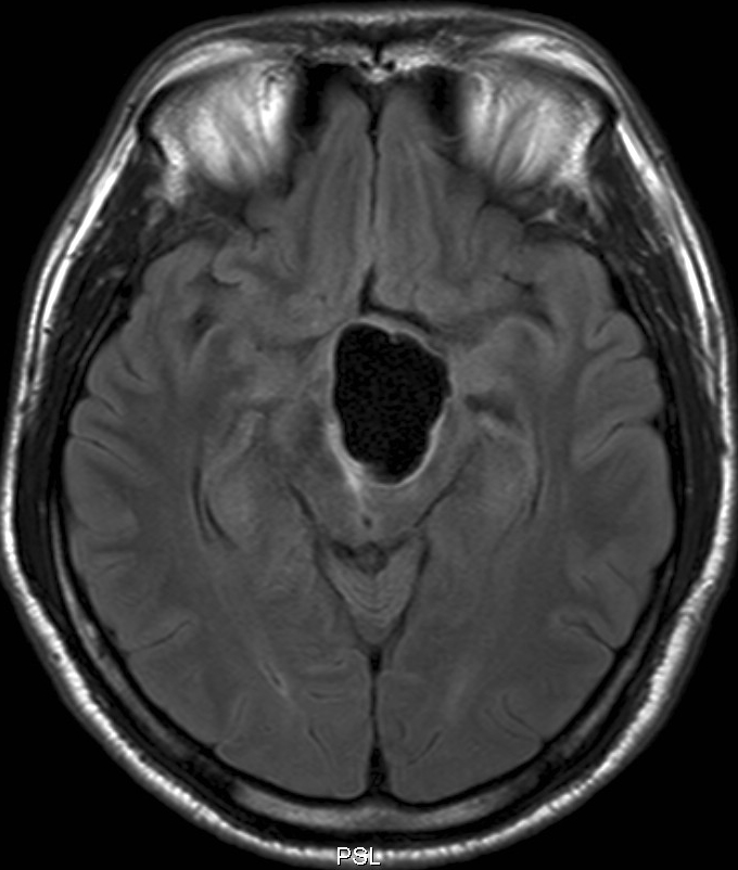
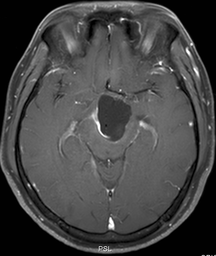
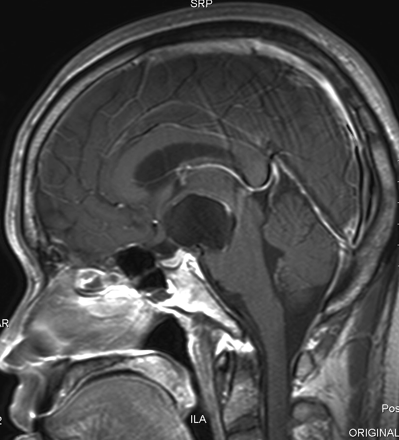
| Discussion | 1. Cysticercosis, cisternal type 2. 영상 소견 약 3cm 크기의 lobulated cystic mass가 suprasellar 및 interpeduncular cistern에서 관찰되며 우측 wall에서 thin rim enhancement를 보인다. Scolex를 시사하는 mural nodule은 보이지 않는다. 3. 고찰 Cysticercosis는 대부분 parenchymal type이나 가끔 cistern이나 ventricle에 발생한다. Cistern이나 ventricle에 발생하는 경우 cisticercus cellulosae 및 cisticercus racemosus라는 두가지 larval form에 의해 감염될 수 있다. 후자의 경우 전자와 달리 scolex를 형성하지 않아 크기가 작을 때는 주변 CSF 신호와 구별이 어려워 진단하기 어렵다. 대신 작은 cystic lesion 들이 서로 뭉쳐서 (conglomerated) 관찰되는 경향이 있고, 뇌막에 염증을 초래하여 leptomeningeal enhancement와 hydrocephalus를 초래할 수 있으나 본 증례에서는 동반되지 않았다. 4. 참고 문헌 - Chang KH, et al. MRI of CNS parasitic disease. JMRI 1998;8:297-307 |
| Correct answer |
Correct AnswerSemi-Correct Answer |