HOME > Weekly Case > Weekly Case
Weekly Case
종결
Case No. 010
0000-00-00~0000-00-00
출제자 : 관리자
Hit : 531
| Period | 0000-00-00~0000-00-00 |
|---|---|
| Diagnosis | |
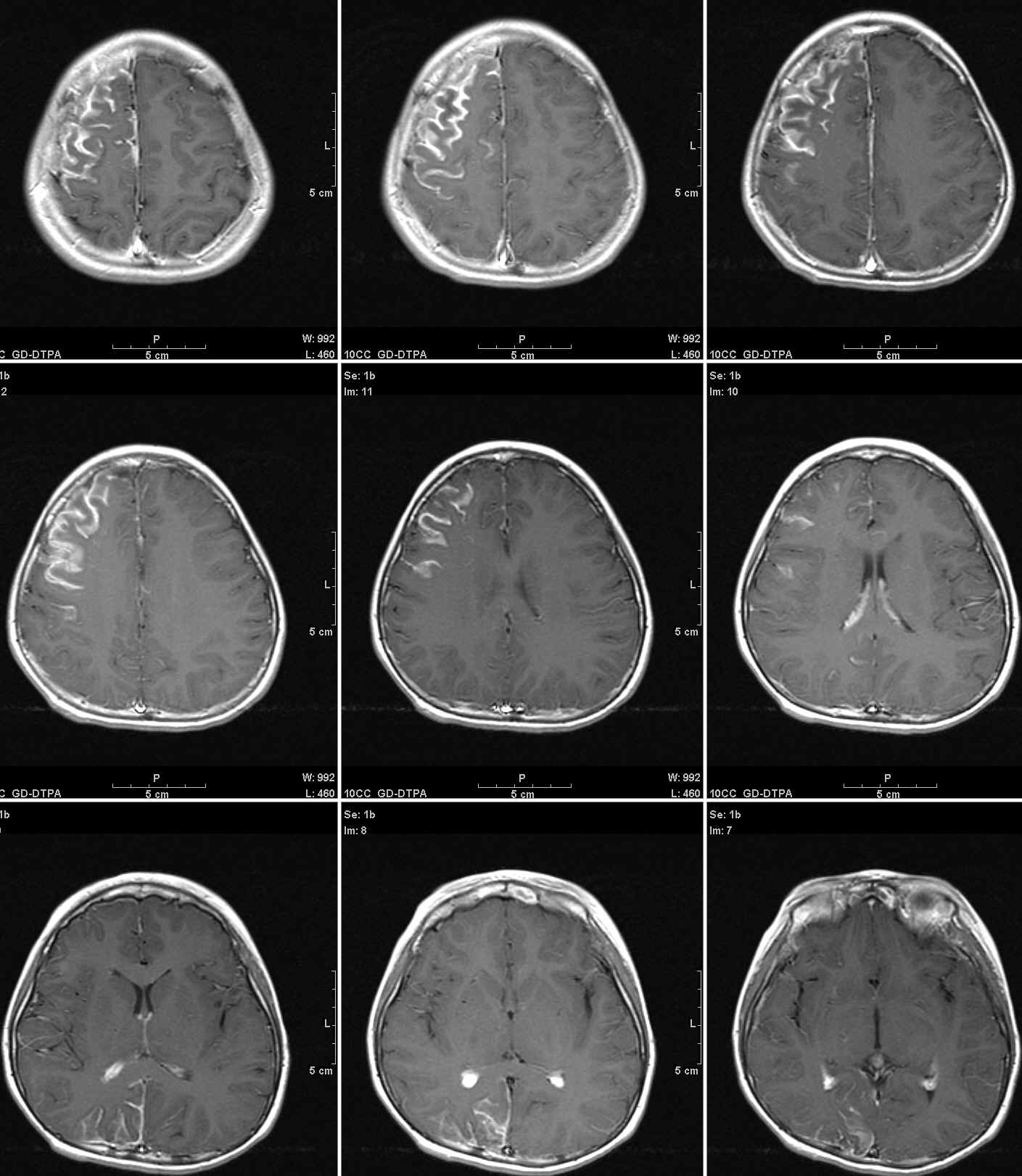
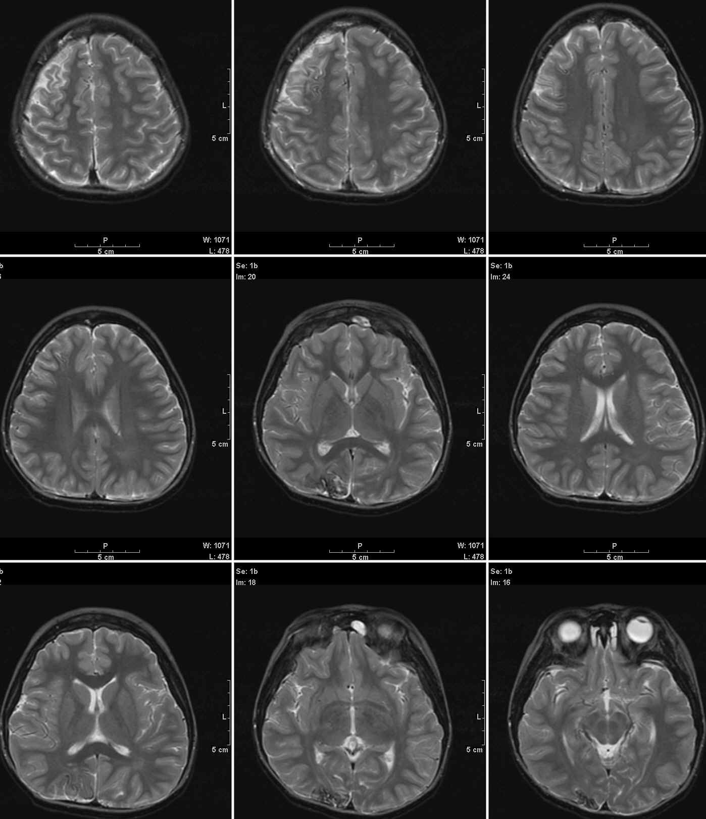
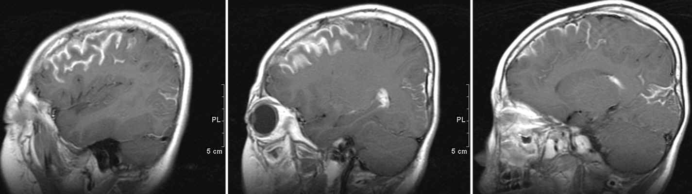
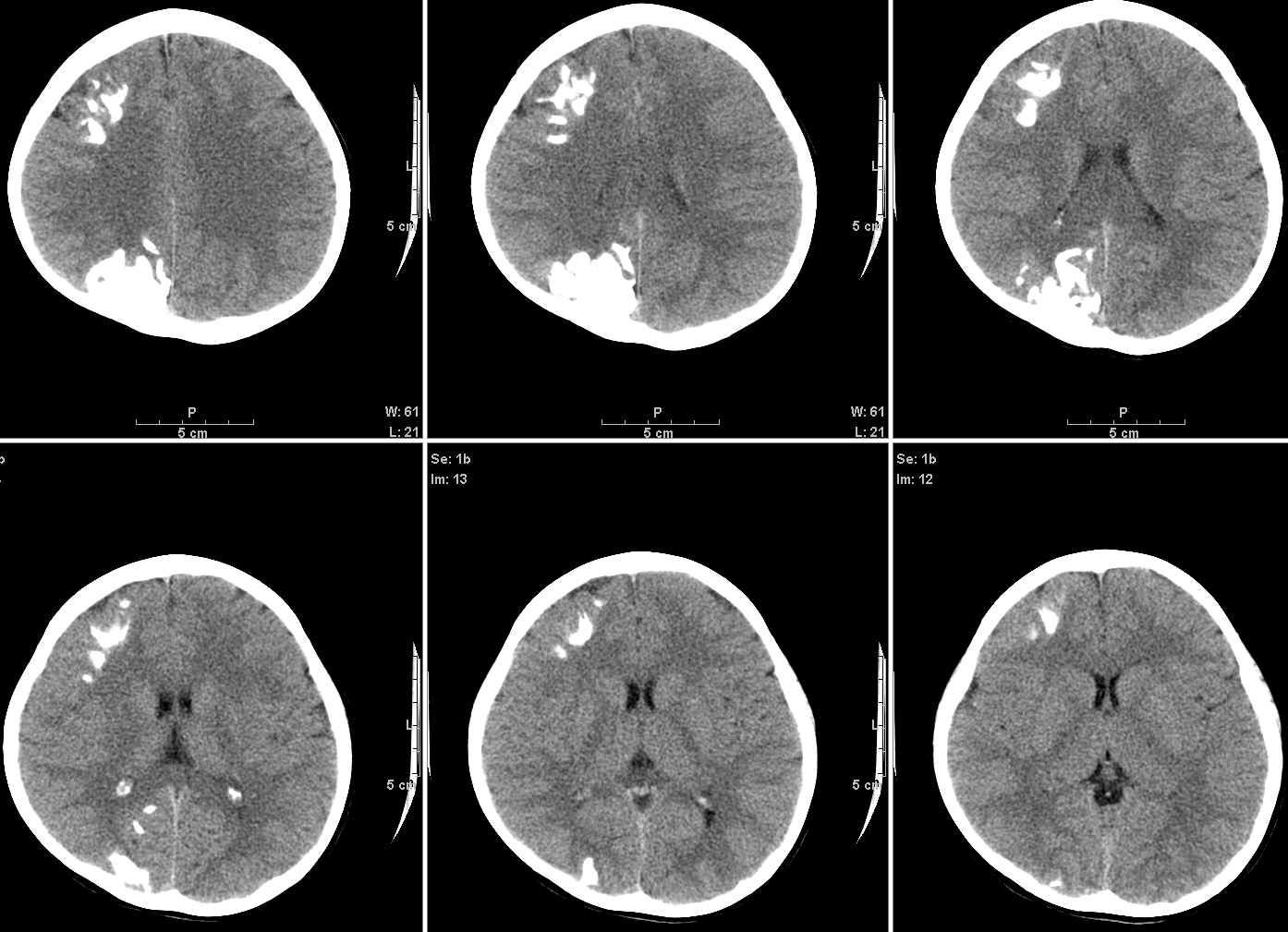
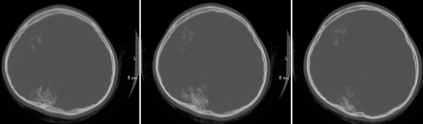
| Clinical information | 7세 소아, Seizure로 영상 시행. 가장 가능성이 높은 진단은? |
| Discussion | 정답: Encephalotrigeminal angiomatosis (Sturge-Weber Syndrome). CNS (Brain and eye)와 face, head의 soft tissue를 침범하는 vascular malformation. Soft tissue의 경우 5번 뇌신경이 지배하는 부위와 일치함. Clinical triad는 seizures, mental handicap, port-wine nevus of face. 그 외 nevus와 동측의 cerebral hemiatrophy, facial hemihypertrophy, 반대측의 somatic hemiatrophy가 있을 수 있슴. 안구는 buphthalmos, choroidal angioma, episcleral telangiectasia가 흔함. Brain은 거의 대부분 involve하며 occipital lobe을 주로 침범하고 temporal, parietal lobe도 침범함. 병의 원인으로 설명되는 이론으로서 persistent primitive vascular plexus가 있슴. 정상적으로 발달하여야 하는 cortical bridging vein들이 형성되지 않아 primitive vascular plexus가 계속 남아 있게 되며, cerebral capillary outflow에 장애가 생기고 subarachnoid space의 작은 혈관으로 drain됨. 출생 후 산소요구량이 증가하여 CBF가 증가하게 되고 venous drainage가 점점 심각하게 감소하여 venous hypertension을 초래함. 점점 진행되면 BBB 손상이 오고 ischemia에 빠져 나중에 dystrophic calcification이 생긴다고 함. 점점 진행하므로 hemispherectomy가 도움이 됨 (특히 5세 이전). 영상소견은, cerebral cortical atrophy, gyriform cerebral calcification, compensatory ventricular enlargement, angiomatous enlargement of ipsilateral choroid plexus, calvarial hemihypertrophy, superficial gyriform enhancement (slow flow vessel and/or BBB injury). |
| Correct answer |
Correct AnswerSemi-Correct Answer |